What does your lumbar MRI mean? How important is the bulging disc or the disc degeneration? Do you need to be worried?
An MRI of your spine will inevitably show something “wrong.” A herniated disc, bulging disc, protruding disc, disc degeneration, annual tear, etc. The list is long.
Patients and healthcare providers alike focus and fixate on lumbar MRI findings. As a result the treatment of lower back pain involves chasing the pain. The structures said to be “abnormal” or “pathologic” on MRI are singled out and then an attempt is made to “fix” them.
The result is usually in an incredible amount of time and money spent seeking out specialists. Physical therapists, chiropractors, pain interventionist doctors, pain management doctors, surgeons, massage therapists, acupuncturists, etc. One or several of these specialists may offer an intervention or approach that works, until the pain comes back. If it ever completely goes away. Sometimes nothing works.
If the pain doesn’t eventually go away frustration, confusion, and even despair set in.
This process spans months to years. Often times another lumbar MRI is ordered to see if something was missed, or if something new is going on. Especially when the pain is worse or has been present for an extended period.
And the cycle repeats itself. Unfortunately the focus on lumbar imaging is misguided. The technology is undoubtedly impressive. With a lumbar MRI you can literally look at the discs, joints, muscles, nerve roots, and vertebrae. It appears to be the perfect tool to diagnose lower back pain. Except it’s not.
In fact, the MRI of your lower back isn’t too important. Unless it shows a tumor, fracture, significant spinal cord compression, or significant nerve root compression that’s correlated with clinical findings. 90% of the time these types of findings are absent.
How could a lumbar MRI not matter?
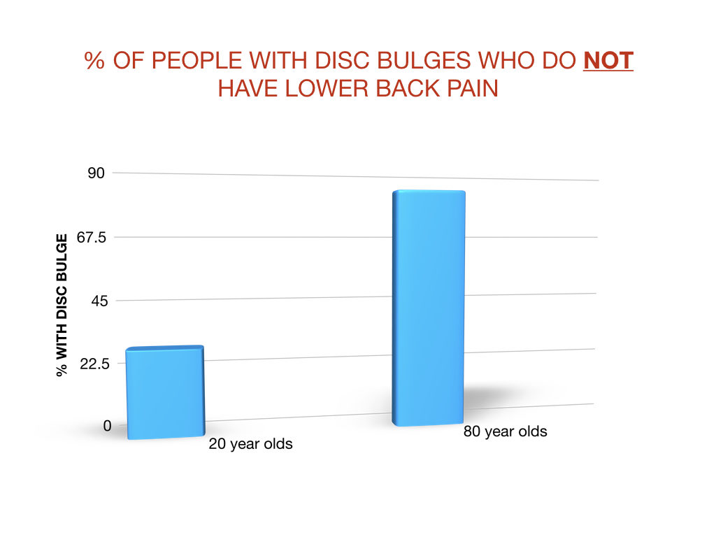
The medical literature is consistent and clear. Many of the findings on lumbar MRI are incidental. Basically people with lower back pain AND people without lower back pain both have disc bulges, disc degeneration, herniated discs, annular fissures, and protruded discs.
In addition, as a person ages the incidence of bulging discs, disc degeneration, and annular fissures increases. IN PEOPLE WITHOUT low back pain. Check out the research.
Systematic literature review of imaging features of spinal degeneration in asymptomatic populations.
37% of 20 year olds have disc degeneration. 96% of 80 year olds have disc degeneration.
19% of 20 year olds have annular fissures. 29% of 80 year olds have annular fissures.
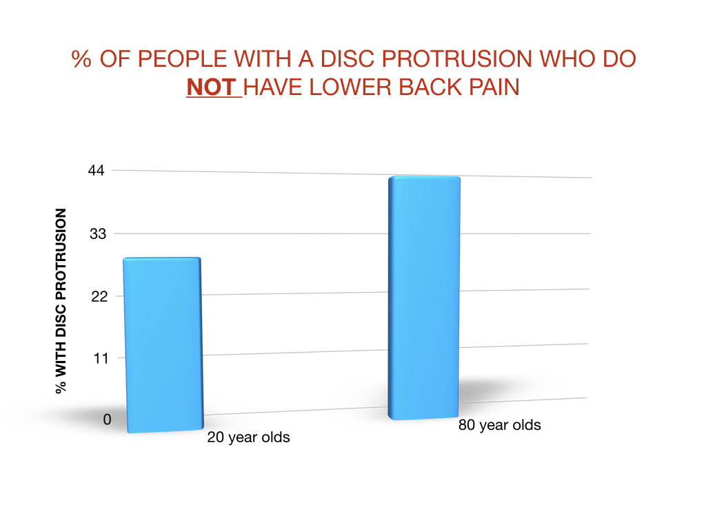
By the time a person is in their twenties it is common to have a disc bulge, disc degeneration, disc protrusion, or annual fissures. WITHOUT LOWER BACK PAIN.
Then, as a person ages it becomes more and more likely that these types of findings will show up on a lumbar MRI. Not because something is wrong with the low back.
Because these types of changes are part of the NORMAL aging process.
Think of disc disc degeneration, disc bulges, and annual fissures as wrinkles on the inside.
Most people don’t love their wrinkles but understand that they’re part of getting older. People understand that they’re not sick and that nothing is wrong with them if they have wrinkles.
Lots of MRI findings we blame back pain on are the same as wrinkles. A simple part of the way our bodies change with time and age.
What about MRI findings in people WITH low back pain?
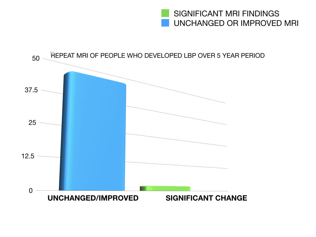
Incredibly when people without lower back pain develop lower back pain more than 80% of them have unchanged or IMPROVED MRI findings.
This study took 200 people WITHOUT lower back pain and followed them for five years. MRIs were taken before they had pain. Then taken again if they developed lower back pain. Even with the onset of lower back pain new MRIs did not show any “smoking guns” in 84% of the cases.
Are first-time episodes of serious LBP associated with new MRI findings?
So how important are lumbar MRI findings?
Clearly not important enough to make them the entire focus of treatment. This sounds counterintuitive, but it’s the truth.
MRI findings that we consider abnormal in people with low back pain are found in people WITHOUT low back pain. And these findings increase in incidence with age.
They can be viewed more as normal, age related changes. Not problems that must be fixed.
Yes, the bulging disc can cause back pain. But don’t focus and fixate on the bulging disc. Focus on doing what has been proven time and again to work.
Exercise. Proven, Powerful Exercises for Lower Back Pain outlines a four week exercise program. The exercises outlined are simple and effective. They can be completed at home, without any special equipment.
Medical and rehabilitation research show that focusing on exercise and function results in less pain and better quality of life compared to chasing the pain.
FAQs
The process itself usually takes right around 30 minutes. Although it may take up to 60 minutes.
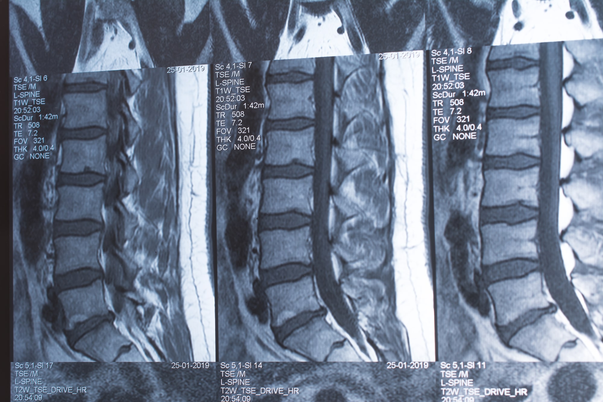
A radiologist will read your lumbar MRI and report pertinent findings. The lumbar MRI will focus on the discs, nerves, spinal cord, muscles, and joints of the lumbar spine. As most people, even physicians, don’t read lumbar MRIs daily the radiologist, back specialist, or surgeon is depended on to interpret the MRI. It’s difficult for the layperson to look at an MRI and know what they’re looking at.
An MRI of the lumbar spine shows the discs, spinal cord, nerves, joints, and muscles of the lumbar spine. A lumbar MRI can be used to determine if lower back pain is related to changes in the discs, nerves, spinal cord, or joints of the lumbar spine. An MRI of the lumbar spine is also used to rule out tumors and fractures that could be causing lower back pain.
An MRI report of the lumbar spine will usually mention disc space narrowing, bulging discs, herniated discs, disc protrusion, annular fissures, nerve root abutment, facet joint arthropathy, vertebral endplate changes, etc. A host of other changes may be mentioned as well, depending on the MRI. Those mentioned are especially common findings. Many of these findings are incidental and in fact normal. The findings on an MRI report need to be matched with specific patient symptoms.
Reading an MRI report often alarms the person reading it because of all the medical terminology and the fact that many of the terms used sound ominous. It’s important to remember that most people without lower back pain have the same findings on MRI when compared to people with lower back pain.
